
Subcellular Nanodomains in Arrhythmias
Alexey Glukhov, PhD, leads research that aims to determine the cellular and molecular mechanisms underlying normal electrical activity and dysfunction of the sinoatrial node, the heart’s natural pacemaker. This work may lead to novel strategies for atrial fibrillation treatment and risk stratification.
Mechanisms of Cardiac Remodeling and Arrhythmogenesis
Dr. Glukhov's lab investigates the cellular and molecular mechanisms of cardiac remodeling and arrhythmogenesis, with the goal of identifying novel diagnostic tools and therapeutic targets. His team focuses on elucidating the functionality of subcellular nanodomains and their role in regulation of proteins responsible for normal and pathophysiological electromechanical activity of the heart.
His laboratory is part of the Cellular and Molecular Arrhythmia Research Program (CMARP) and the Cardiovascular Research Center (CVRC).
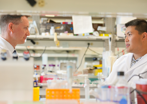
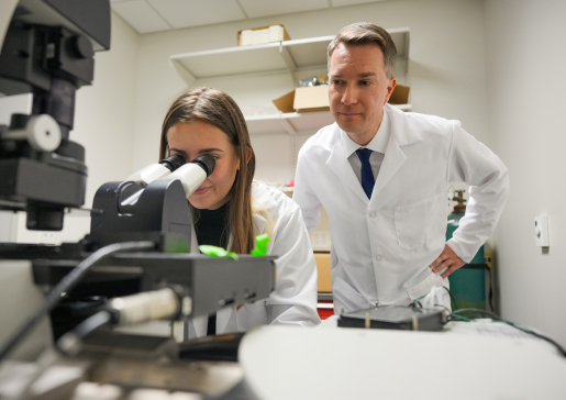
Research Team

Scientist I
Scientist I

Research Associate
Students
- Jenson Aaron
- Pavan Yilayavilli
Active Projects
- Microdomain-Specific Localization of Functional Ion Channels in Cardiomyocytes
It has recently become evident that discrete clusters of different ion channels and regulatory receptors are present in the sarcolemma, where they form an interacting network and work together as a part of a macro-molecular signaling complex which in turn allows the specificity, reliability and accuracy of the autonomic modulation of the excitation–contraction processes by a variety of neurohormonal pathways.
Disruption in subcellular targeting of ion channels and associated signaling proteins may contribute to the pathophysiology of a variety of cardiac diseases, including heart failure and certain arrhythmias.
Our laboratory is particularly interested in understanding the functionality of distinct subcellular microdomains in cardiac myocytes, such as transversal (t)-tubules, caveolae, and various costamers, and their functional role in the accumulation, neuro-hormonal and mechanical regulation of proteins responsible for normal and pathophysiological electrical activity of the heart.
- Functional Microdomains in the Sinoatrial Node
Our group developed a conceptually innovative approach to study sinoatrial node dysfunction through functional protein-protein interactions within discrete nanodomains.
In particular, we focus our research on specialized cardiomyocyte membrane structures, namely caveolae, submicroscopic plasma membrane pits, that organize sinoatrial node pacemaking through complex spatiotemporal cAMP-dependent and Ca2+-mediated crosstalk between sarcoplasmic reticulum and sarcolemmal membrane proteins within the so-called coupled-clock pacemaker system that support a stable, regular sinus rhythm.
This pioneering research introduces a novel concept of electrophysiological changes resulting from physical alterations in the subcellular localization of and interactions between signaling complexes following structural remodeling.
- Mechano-Electrical Signal Transduction in Pathophysiology of Atrial Fibrillation
Atrial fibrillation is the most common cardiac rhythm disorder worldwide and often occurs in the setting of hypertension and associated atrial dilatation with pathologically increased cardiomyocyte stretch. In the setting of atrial dilatation, mechano-electric feedback has been linked to the development of ectopic beats that trigger paroxysmal atrial fibrillation originating mainly from pulmonary veins as well as from non-pulmonary vein foci.
Our group identified caveolae-mediated activation of mechanosensitive volume-regulated chloride channels ICl,swell as a critical cause of pulmonary vein ectopic beats that can initiate atrial arrhythmias. We demonstrated that downregulation of caveolae structures during chronic hemodynamic overload of the atria facilitates activation of ICl,swell and increases sensitivity of pulmonary veins to stretch.
These findings provide conceptual innovation on cardiac mechanosensing and form a mechanistic basis for development of novel and effective therapeutic approaches targeted to treat stretch-induced atrial arrhythmogenesis via preventing the degradation or promoting the restoration of cardiac cytoarchitecture.
- The Wisconsin Atrial Fibrillation Initiative: Integrating Novel Technologies to Tackle a Pervasive Disease
In collaboration with Dr. Alejandro Roldán Alzate (Departments of Mechanical Engineering and Radiology) and Dr. Matthew Kalscheur (Department of Medicine), we have shown, in both animal model and patients with atrial fibrillation, that pathological stretch heterogeneously affects atrial myocardium creating distinct “vulnerable” regions of pathophysiological remodeling and arrhythmogenesis.
By utilizing 4D flow magnetic resonance imaging (MRI) combined with computational fluid dynamics, our collaborative team propose a highly innovative approach for identifying regions of stretch-induced myocardial remodeling and proarrhythmic ectopy as areas of local hypokinetic behavior.
This will allow the development of a much-needed non-invasive tool for the assessment of the degree of electro-anatomical remodeling of patient atria and prediction criteria for atrial fibrillation risk stratification in vulnerable cohorts of patients.
Funding Support
Dr. Glukhov’s research is funded by the National Institute of Health and the American Heart Association.