Study identifies how Candida albicans creates drug-resistant biofilms
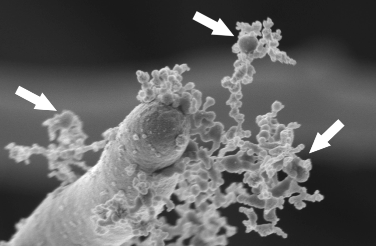
Image (above): A scanning electron micrograph shows Candida albicans yeast (elongated structure at left) building an antifungal drug-resistant biofilm structure by secreting small, spherical extracellular vesicles (arrows) that deliver payloads of biofilm polymers to the extracellular matrix.
The pathogenic yeast Candida albicans has a clinically frustrating habit: when it colonizes the surfaces of indwelling medical devices such as catheters, joint protheses, and mechanical heart valves, it assembles into a tough, durable structure. This structure, called a biofilm, enables immune system evasion and is nearly impervious to antifungal drugs.
Biofilms can be deadly, leading to bloodstream infections and invasive systemic infections of tissues and organs. Catheter microbial biofilms are estimated to be responsible for 100,000 deaths and have an economic impact of $6.5B annually in the United States alone.
Understanding how a drug-resistant biological barrier forms is the first step to disrupting it. That’s the premise of recent research led by David Andes, MD (pictured at right), professor and head, Division of Infectious Disease in the Department of Medicine.
Previously, members of the Andes laboratory discovered that a key step in the formation of C. albicans biofilms is the yeast species' ability to make a protective carbohydrate substance consisting of a mannan-glucan carbohydrate complex.
They then turned attention to how this material is delivered to the exterior of the cell to serve as biofilm building materials.
Using electron microscopy, the scientists spotted a clue: small spheres emanating from C. albicans cells growing in biofilms. The spheres were less than 100 nm in diameter (for comparison, a human hair is about 75,000 nm thick). This size range suggested that they could be extracellular vesicles, which are balloon-like bubbles found throughout the microbial world that often serve as delivery vehicles to transport materials that are protectively surrounded with a fatty lipid bilayer.
Further analyses using yeast mutant strains that are unable to produce extracellular vesicles effectively showed that the spheres were, in fact, extracellular vesicles.
“We were surprised to find that vesicles were the mechanism of delivery of the biofilm protective shield,” said Dr. Andes. “Our main challenge was – and continues to be - determining which components of the vesicle cargo are responsible for protection.”
Additional experiments showed that biofilm-associated extracellular vesicles were distinct in size from vesicles produced by free-living C. albicans cells. Biochemical analyses also showed that the cargo of biofilm extracellular vesicles largely correspond to materials found in mature biofilms.
What does this mean for the development of new treatments for C. albicans infections? In a word: hope. Several mutant strains with limited capability to produce extracellular vesicles created biofilms that were useless to the yeast in terms of protection against the antifungal drug fluconazole. When researchers added normal biofilm extracellular vesicles to these strains, they became drug-resistant once again.
“Our preliminary data suggest targeting key enzymes delivered in the vesicles may serve a drug targets that will ‘unprotect’ the fungus from other common antifungal drugs,” said Dr. Andes. “In addition, the unique vesicle cargo may also provide a unique biofilm signature that may be used as a biofilm diagnostic target.”
The research article received attention from other outlets, including Nature Reviews Microbiology. Implications of the findings are far-reaching, explains Dr. Andes.
“Many of my biofilm and fungal colleagues are now wondering if virulence and pathogenesis factors that they study may also be delivered by this process.”
Dr. Andes holds the William A. Craig Endowed Professorship. Additional authors of the research article are Robert Zarnowski, PhD, associate scientist; Hiram Sanchez, assistant researcher; Antonio S. Covelli, research specialist; Eddie Dominguez, graduate student; and Kaitlin F. Mitchell, graduate student, all of University of Wisconsin-Madison; Christian Heiss and Parastoo Azadi of the Complex Carbohydrate Research Center at the University of Georgia; Aaron Mitchell of Carnegie Mellon University; Anna Jaromin of University of Wroclaw in Poland; and Jörg Bernhardt of Ernst Moritz Arndt University Greifswald in Germany.
Resources:
- Zarnowski R, Sanchez H, Covelli AS, Dominguez E, Jaromin A, Bernhardt J, Mitchell KF, Heiss C, Azadi P, Mitchell A, Andes DR. 2018. Candida albicans biofilm-induced vesicles confer drug resistance through matrix biogenesis. PLoS Biol. 16(10):e2006872.
- Du Toit A. 2018. Sharing with your community. Nat Rev Microbiol. (12):718-719.
- "Funding awarded to Dr. David Andes for research on medical device biofilms," Department of Medicine, July 23, 2018